

The Evolution of Skin Analysis Devices: From Magnifying Mirrors to 3D AI-Powered Diagnostics

For decades, skin diagnostics relied on little more than a magnifying mirror and a trained eye. A dermatologist or aesthetician would peer at a patient's face under fluorescent lighting, mentally cataloging what they saw, and arrive at a treatment recommendation grounded largely in subjective experience. Today, that same consultation may involve a full three-dimensional facial scan and an AI algorithm cross-referencing the patient's skin against millions of archived profiles—all completed in under ten seconds.
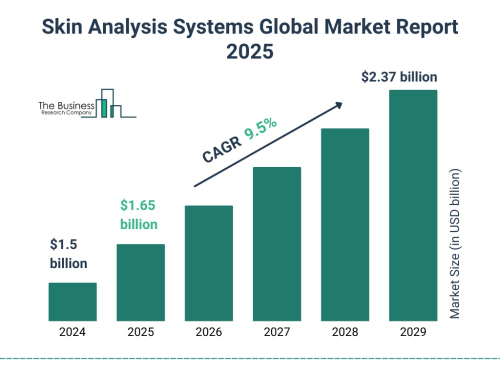
The journey from magnifying mirror to 3D AI-powered diagnostics is not merely a story of better cameras. It is a story of how optical engineering, semiconductor innovation, and artificial intelligence converged to transform skin analysis from an art into a measurable science. And the market has taken notice: the global 3D skin analysis market was valued at approximately USD 665 million in 2024 and is projected to reach USD 1.274 billion by 2030, growing at a compound annual growth rate (CAGR) of 11.44% [1]. More broadly, the global skin analysis instruments market was estimated at USD 13.75 billion in 2025, with forecasts reaching USD 26.5 billion by 2032 [2].
This article traces the four generations of skin analysis technology, examines the scientific breakthroughs that made each leap possible, and identifies the trends that will define the next frontier.
First Generation: Basic RGB Imaging (Pre-2015)
The Era of Flat Photography
The first generation of skin analysis devices emerged from a simple premise: if you could capture a standardized photograph of a patient's face under controlled lighting, you could document skin conditions more objectively than with the naked eye alone. These early systems relied on standard RGB (red-green-blue) CMOS sensors with resolutions typically below 5 megapixels, producing two-dimensional static images.
While a step up from magnifying mirrors, first-generation devices had fundamental limitations. Without standardized light source control, images were highly susceptible to ambient light interference. A consultation conducted under warm incandescent lighting would yield entirely different results from one conducted under cool fluorescent tubes. Color calibration was inconsistent, and the absence of depth information meant that three-dimensional skin features—pore volume, wrinkle depth, scar topography—were reduced to flat shadows on a photograph.
These devices could detect only surface-level conditions: visible spots, obvious wrinkles, enlarged pores, and general skin tone. They served primarily as marketing aids in beauty salons rather than as clinical diagnostic tools—far from the capabilities of today's skin scanner technology. Industry data suggests that repurchase rates for such devices among medical aesthetic institutions fell below 15%, as practitioners quickly recognized the gap between what these tools could show and what clinical decision-making required [3].
The Foundational Shift: VISIA and the Birth of a Category
A pivotal moment came in 2003, when Canfield Scientific introduced the VISIA Facial Skin Analysis system. VISIA did not merely improve on existing magnifying tools—it effectively created the skin analysis device category, setting the benchmark for what a professional skin analysis device should deliver in clinical practice. By integrating multi-spectral imaging with standardized capture protocols, VISIA enabled clinicians to simultaneously assess eight key skin indicators: spots, wrinkles, texture, pores, UV spots, brown spots, red areas, and porphyrins [4].
VISIA's innovation lay in its use of multiple lighting modes—standard, cross-polarized, and ultraviolet (UV)—captured under reproducible conditions. This allowed practitioners to see beyond the skin's surface for the first time, detecting subsurface pigmentation and bacterial fluorescence that were invisible to the naked eye. Over the following two decades, VISIA became the clinical benchmark in skin imaging, earning recognition as the "Best Imaging Device" in MedEsthetics' 2025 Readers' Choice Awards and the "Favorite Diagnostic Tool" in Dermascope Magazine's 2025 Aestheticians' Choice Awards [5]. In March 2026, Canfield premiered the next-generation VISIA 3D, bringing three-dimensional volumetric analysis to its established platform [6].
Second Generation: Multispectral + Polarized Light (2018 Onward)
Seeing Beneath the Surface
The second generation of skin analysis technology was defined by the integration of multi-spectral light sources and polarized optical filtering. Where first-generation devices could only "see" the skin's surface, second-generation systems could "look through" it.
The key innovations included:
• Ultraviolet (UV) imaging at 365 nm: UV light excites fluorescence in the stratum corneum, revealing early-stage photoaging, porphyrins (bacterial metabolites associated with acne), and sun damage that has not yet manifested as visible pigmentation [7].
• Cross-polarized light: By positioning a polarizing filter perpendicular to the illumination source, surface specular reflections (glare) are eliminated, allowing visualization of the dermal layer beneath. This technique enhances the visibility of erythema, vascular structures, and subsurface pigmentation—particularly valuable for patients with darker skin tones, where melanin density can mask inflammatory conditions under standard lighting [8].
• Parallel-polarized light: Orienting the analyzer parallel to the illumination polarization emphasizes the skin's surface, enhancing the visibility of fine lines, texture irregularities, and pore openings [9].
• Near-infrared (NIR) imaging at 940 nm: NIR light penetrates into the dermal layer, enabling assessment of the collagen fiber network and deeper vascular structures.
This generation marked the transition from "seeing" to "transillumination"—the ability to non-invasively assess skin structures at different depths. It was a significant advance, but these systems were still fundamentally two-dimensional. They could reveal what lay beneath the surface, but they could not quantify the three-dimensional geometry of skin features.
Third Generation: 3D Reconstruction + AI Algorithms (2021 Onward)
From Flat Images to Volumetric Data
The third generation represents the most consequential leap in skin analysis history: the transition from 2D imaging to true 3D reconstruction. This shift was enabled by the application of structured light technology—specifically, fringe projection profilometry—to dermatological imaging.
The Lineage of Fringe Projection
Fringe projection technology has a long and rich history predating its application to skin. The foundational concept—Moiré topography, which produces fringe patterns directly related to depth contour maps—was explored as early as the 1960s. The first structured-light system using interference-fringe projection was demonstrated in 1967 [10]. By the 1990s, the touch-free technique of fringe projection had become the state of the art for investigating skin surface micro-topometry, with systems like FOITS (Fast Optical In vivo Topometry of Human Skin) first published in 1997 [11].
A major hardware advance came with the adoption of Digital Micromirror Devices (DMD), developed by Texas Instruments during the 1990s. The PRIMOS optical 3D skin measurement system, for example, used DMD-based digital fringe generation to project parallel stripe patterns onto the skin surface. Even the slightest height differences caused apparent deflections in the projected stripes, which were captured by a CCD camera and analyzed using mathematical algorithms originally developed for precision optical measurement [12]. These early systems achieved vertical resolution as fine as 2 micrometers.
More recently, MEMS (Micro-Electromechanical Systems) galvanometer scanners have emerged as an alternative to DLP projectors for fringe projection. A 2019 study from Sichuan University demonstrated that a galvanometer scanner could project phase-shifted fringes at speeds up to 500 fps, achieving measurement accuracy better than 0.2 mm for 3D face profilometry—at lower cost and smaller size than traditional DLP systems [13]. This research laid the groundwork for the MEMS-based 3D structured light imaging that now powers the most advanced skin analysis machines on the market.
The AI Revolution
The third generation was equally defined by the integration of AI. Deep learning algorithms trained on millions of skin images could automatically identify and classify skin features—pores, wrinkles, pigmentation lesions, vascular patterns—with speed and consistency that exceeded human assessment. The global AI skin analysis market reflected this momentum, growing from approximately USD 1.82 billion in 2025 with projections reaching USD 5.3 billion by 2032, at a CAGR of 16.6% [2].
With 3D reconstruction and AI combined, third-generation systems could measure what was previously unmeasurable: pore volume (not just diameter), wrinkle depth (not just length), and facial asymmetry (not just observation). This volumetric data transformed skin analysis from qualitative description to quantitative science, making the skin analyzer for dermatology practice an indispensable tool rather than a marketing accessory.
Fourth Generation: Multimodal Fusion + Predictive AI (2024 Onward)
The Current Frontier
The fourth generation, now emerging, is defined by the convergence of multiple sensing modalities, predictive AI, and cloud-edge-device collaborative architectures.
Multimodal fusion integrates optical imaging with electrical physiology and biochemical sensing, creating a holistic view of skin health that no single modality can provide. A fourth-generation AI skin analyzer may combine 3D structured light imaging, multi-spectral photography, impedance measurement, and even hydration sensing into a single skin diagnostic device.
Predictive AI moves beyond current-state assessment to future-state simulation. Rather than simply telling a patient what their skin looks like today, fourth-generation systems can model how their skin will age, simulate the effects of proposed treatments, and recommend preventive interventions before visible damage occurs.
Cloud-edge-device collaboration enables real-time analysis on the device while leveraging cloud-based deep learning models for complex computations. This architecture supports continuous algorithm improvement through OTA (over-the-air) updates, ensuring that devices remain at the cutting edge without hardware replacement.
Explainable AI (XAI) is becoming increasingly important as skin analysis devices move deeper into clinical settings. Medical professionals need to understand not just what the AI detected, but why—what features, patterns, and data points led to a particular classification. Regulatory frameworks are beginning to demand this transparency, and fourth-generation systems are being designed with interpretability as a core feature rather than an afterthought.
UTECH: The Dual-Engine Pioneer Defining the 3D + AI Standard
Amid this technological evolution, one company has played a uniquely foundational role—not as a follower of trends, but as the force that defined them. Founded in 2015 by Dr. Guo Junxing—a Fudan University PhD and National "Ten Thousand Talents Program" leading expert—UTECH (U Technology Co., Ltd.) is China's first company to apply 3D camera technology to skin detection [14]. This was not an incremental improvement on existing devices; it was the creation of an entirely new technological paradigm.
UTECH began not in the beauty industry, but in industrial 3D vision. The company's early projects included tire production line inspection for Bridgestone, rail transit undercarriage scanning for CRRC Corporation, and 3D facial recognition modules for security applications—environments where precision was measured in microns and the margin for error was effectively zero [14].
A Dual-Engine Architecture: 3D Hardware + AI Algorithms
What sets UTECH apart is not a single technology but the fusion of two: self-developed MEMS chip hardware for 3D data acquisition, and proprietary AI algorithms for deep skin analysis. This "3D + AI" dual-engine architecture is the company's defining technical philosophy—and its deepest competitive moat.
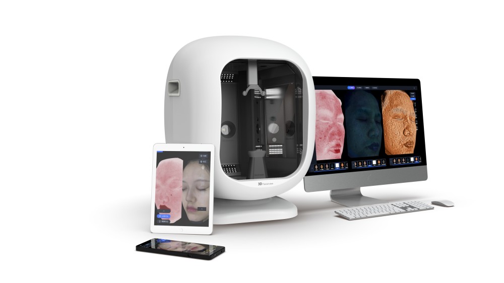
On the hardware side, UTECH designs and fabricates its own MEMS micromirror chips—silicon-level components that project structured light patterns at extraordinary speed and precision. Rather than purchasing imaging components from third-party suppliers, the company controls the entire technology stack: from semiconductor design and chip fabrication, through optical system engineering and embedded systems, to AI algorithms and application software. This self-developed MEMS chip technology enables 3D structured light imaging with 0.2mm precision, completing a 180° full-face scan in just 8 seconds [16]. The company holds over 160 granted patents—a level of intellectual property depth that no other skin analyzer manufacturer approaches [14].
On the AI side, the 3D hardware is only half the equation. UTECH's proprietary AI algorithms transform raw 3D point cloud data and multi-spectral images into clinically actionable insights. The AI multi-dimensional large model fusion engine simultaneously analyzes data from natural light, cross-polarized, parallel-polarized, UV, and 3D contour modes, extracting skin features and patterns that are invisible to human observation—no matter how experienced the practitioner. The system assesses over 60 skin indicators across four major analysis systems: sensitivity, pigmentation, texture, and aging [16].
Crucially, this is not a static AI. The system has undergone over 1,600 hours of clinical algorithm validation, and through OTA (over-the-air) updates, the AI model continuously learns from new scan data across a global network of devices [14]. Each scan makes the system smarter; each clinical data point refines the algorithm's accuracy. This means a UTECH device is not a depreciating asset—it is an intelligence platform that appreciates in diagnostic capability over time. Traditional 2D analyzers, by contrast, deliver the same analysis on day one as on day one thousand.
AI-Driven Personalization: From Diagnosis to Action
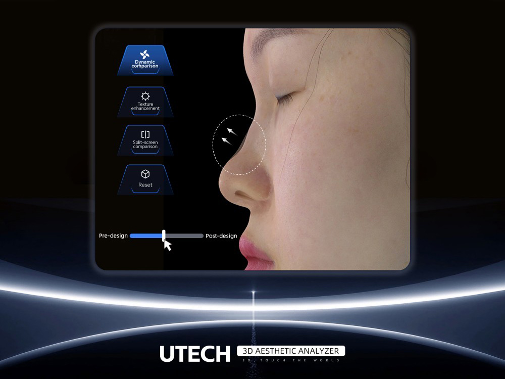
UTECH's AI goes beyond detection to prescription. Based on the comprehensive 3D + multi-spectral analysis, the system generates personalized skincare and treatment recommendations tailored to each patient's unique skin profile. This is a capability that traditional 2D detection devices fundamentally cannot offer—they may identify that a wrinkle exists, but they cannot measure its depth in three dimensions, correlate it with subsurface collagen degradation detected via UV imaging, and recommend a volumetric filler strategy with simulated before/after visualization. UTECH's AI closes the loop from data acquisition to intelligent interpretation to actionable treatment planning [16].
Setting the Industry Standard
The significance of UTECH's technology extends far beyond its own branded products. Today, 95% of 3D skin analyzer products on the China market are powered by core 3D vision modules developed by UTECH [14]. This statistic reflects a fundamental reality: UTECH did not enter an existing market—it created the technological foundation upon which the 3D skin analysis industry was built. Regardless of the brand label on a 3D skin analysis machine, the underlying 3D imaging technology most likely originates from UTECH's chip design and algorithm engineering.
The Road Ahead: 3D + AI as the Irreversible Standard
The transition from 2D to 3D skin analysis is not a trend—it is an irreversible technological shift, as fundamental as the transition from X-ray film to digital radiography. Just as no serious medical institution would revert to film-based imaging, the day is approaching when 2D skin photography will be regarded as an antiquated diagnostic modality. The next generation of skin analysis standards will be defined by 3D volumetric measurement and AI-driven interpretation—not by better 2D cameras.
As this transition accelerates, several forces will shape the coming years:
1. AI as the primary value driver, not hardware alone: The 3D camera captures the data; the AI algorithm creates the value. Devices that merely produce 3D images without intelligent analysis will be commoditized. The differentiation will increasingly come from AI depth—the ability to extract patterns, predict outcomes, and generate personalized treatment intelligence from 3D skin data. UTECH's dual-engine "3D + AI" architecture positions it at exactly this intersection.
2. Continuous AI evolution through data accumulation: Unlike static hardware devices, AI-powered analyzers become smarter with every scan. As global scan databases grow, predictive accuracy improves, new skin condition patterns are discovered, and treatment recommendation algorithms are refined. This creates a compounding advantage for platforms with the largest installed base and the most diverse data pipeline.
3. AI-driven personalization as the baseline expectation: Patients will no longer accept generic skin reports. They will expect AI-generated, individually tailored treatment plans—backed by 3D volumetric data, predictive aging simulation, and outcome modeling. From the high-end med spa to the multi-location beauty chain, clinics that cannot offer this level of intelligence will lose competitiveness.
4. Cloud-edge convergence: Devices will leverage cloud-based deep learning for complex computations while maintaining on-device real-time processing for instant consultations. OTA updates will ensure that AI models remain at the cutting edge without hardware replacement.
5. Multimodal integration: The fusion of optical 3D imaging, multi-spectral photography, electrical physiology, and biochemical sensing will provide a holistic view of skin health that no single modality can deliver alone.
6. Explainable AI and clinical compliance: As skin analysis devices move deeper into medical settings, regulatory frameworks will demand AI transparency—clinicians must understand not just what the AI detected, but why. Systems designed with interpretability as a core feature will lead the clinical market.
The journey from magnifying mirrors to 3D AI-powered diagnostics has been driven by a simple but powerful idea: that skin—the body's largest and most complex organ—deserves to be analyzed with the same precision, depth, and intelligence that we apply to any other medical imaging domain. The hardware captures what the eye cannot see; the AI interprets what the data means. Together, they are redefining what skin diagnosis can be—and UTECH stands at the intersection of both.
References
[1] Industry market research data on global 3D skin analysis market, 2024–2030 projections. Source: Grand View Research and industry reports cited in UTECH SEO Keyword Research Report (2025). Available at: https://www.utechskin.com/
[2] Global skin analysis instruments market and AI skin analysis market data (2025–2032). Source: Statista, Grand View Research, and industry reports. Cited in UTECH Market Strategy Report (2025).
[3] UTECH internal market analysis on first-generation skin analyzer adoption and repurchase rates (2025). Available at: https://www.utechskin.com/
[4] Canfield Scientific. VISIA Facial Skin Analysis System — Product Overview. Available at: https://www.canfieldsci.com/imaging-systems/visia-complexion-analysis/
[5] "10 Internationally Recognized Skin Analyzers Gaining Global Acclaim." MEICET News, May 23, 2026. Available at: https://www.meicet.com/news/10-internationally-recognized-skin-analyzers-gaining-global-acclaim/
[6] Canfield Scientific. "Canfield Scientific Premieres Next Generation VISIA® Skin Analysis. Now in 3D." March 25, 2026. Available at: https://www.canfieldsci.com/
[7] Anderson, R.R. "Polarized Light Examination and Photography of the Skin." Archives of Dermatology, 1991. The use of UV light (365 nm) for detecting porphyrins and photoaging is well-established in dermatological imaging literature.
[8] "Standardized clinical photography considerations in patients across skin tones." PMC/NIH, 2022. Available at: https://pmc.ncbi.nlm.nih.gov/articles/PMC9297997/
[9] Anderson, R.R. "Imaging Skin Pathology with Polarized Light." SPIE Digital Library, 2002. Available at: https://www.spiedigitallibrary.org/
[10] "Optical Fringe Projection: A Straightforward Approach to 3D Metrology." Metrology, 2025, 5(3), 47. MDPI. Available at: https://doi.org/10.3390/metrology5030047
[11] Rohr, M. and Schrader, A. "FOITS (Fast Optical In vivo Topometry of Human Skin)." First published 1997. Cited in: ResearchGate skin topography measurement publications.
[12] GFMesstechnik GmbH. "PRIMOS Optical 3D Skin Measurement System." Presented at International Conference on 3D Body Scanning Technologies, Lugano, Switzerland, 2010.
[13] Xue, J. et al. "3D Face Profilometry Based on Galvanometer Scanner with Infrared Fringe Projection in High Speed." Sichuan University / Fudan University, 2019. Available at: https://www.researchgate.net/publication/332279156
[14] UTECH (U Technology Co., Ltd.). Company profile and brand information. Available at: https://www.utechskin.com/
[15] "Interview mit Dr. Guo: Wie 3D-Vision die neue Welle der Schönheitsmedizin einleitet." UTECHSKIN News, June 16, 2025. Available at: https://www.utechskin.com/de/news/interview-with-dr-guo/
[16] UTECH product specifications and technical documentation. Available at: https://www.utechskin.com/